Share this resource or email it to a friend!
The Anatomy of Feline Eyes
To better understand the development and function of kittens’ eyes, it helps to be familiar with the eye’s anatomy.
- The orbit is the bony cavity within the skull in which the eyeballs are situated. The orbit also contains muscles, nerves, blood vessels and the structures that produce and drain tears. The feline orbit is relatively deep, and, even though it is not completely formed of bone, it provides protection for the eye.
- The sclera, also known as the white of the eye, is the opaque, tough and protective outer layer of the eye; it maintains the eyeball’s shape to support internal structures and also serves as an attachment site for extraocular muscles.
- The conjunctiva is a thin mucous membrane that covers the sclera. This membrane is a layer of epithelial cells with mucus-secreting cells, which covers the eye and lines the eyelids. In healthy felines, the conjunctiva is not readily visible and is a pale, pink color.
- The nictitating membrane, or third eyelid, is in the inner corner of the eye and provides extra protection by removing debris from the eye’s surface and redistributing tears over the cornea. The nictitating membrane is usually not visible in healthy eyes unless the cat is relaxed, sleeping or blinking, at which time the third eyelid moves across the eye’s surface.
- The cornea is the transparent dome on the front part of the eye that covers the iris, pupil and anterior chamber (the space between the cornea and iris). The cornea not only protects the front of the eye, but also helps focus light on the retina at the back of the eye.
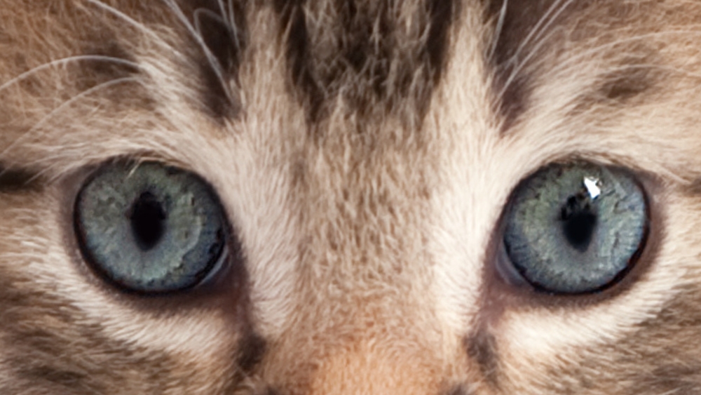
- The iris is the circular, colored area of the eye that controls the amount of light that enters the eye by making the pupil larger or smaller.
- The pupil is the black area in the center of the eye. A cat’s pupil has an elliptical form and is in a vertical orientation. When the sphincter muscles of the iris contract, as in a bright environment, the pupil takes the shape of a narrow slit.
- The lens, which sits behind the iris, is a small, translucent structure that adjusts its shape as needed to focus incoming rays of light on the retina.
- The retina is a light-sensitive tissue that lines the interior surface of the eye. When the retina receives light impulses that have passed through the lens, they are instantaneously transmitted to the brain as visual information via the optic nerve, which is attached to the back of the eye. The retina contains photoreceptors, which are the image-forming cone and rod cells.
- The cone cells provide excellent visual sharpness and binocular vision (a type of vision that utilizes frontal facing eyes with overlapping fields of view, thus providing good depth perception). Cone cells are also responsible for color vision, and, although it is unclear whether cats can see colors, and, if so, which ones, experts believe that cats can see blues and yellows.
- The rod cells collect dim light; cats have 6-8 times more rod cells than cones, which gives them excellent night vision.
- The tapetum lucidum is a specialized layer of flat, mirror-like cells found beneath the retina that allows light to bounce off it and back onto the retina. This reflection is what causes the blue or greenish glint to eyes at night.
- The eyelids are thin folds of skin that cover the eye. They blink reflexively to protect the eye and distribute tears.
- The tears contain moisture, which is essential to healthy eyes. Tears are made up of water, mucus and oil; this mixture provides a more protective tear that is slower to evaporate.
- The lacrimal glands, located at the top outer edge of each eye, produce the watery portion of tears.
- The goblet cells, glands in the conjunctiva, produce the mucus portion of tears.
- The meibomian glands, located within the eyelids, produce the oily portion of tears.
- The nasolacrimal ducts, at the edge of the upper and lower eyelids near the nose, allow the tears to drain into the nose.
The Development of Kittens’ Eyes
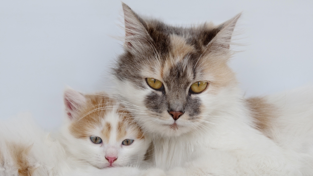
Kittens are born with fused eyelids, which gradually open and are typically fully open between 10-14 days of age. If the eyes remain closed after two weeks, it is advisable to contact the animal shelter, rescue group or your veterinarian. NEVER pry open a kitten’s eyes.
All kittens are born with blue eyes, which will begin to change to their adult eye color between 6-10 weeks of age. During the maturation process, it is common to see various colored flecks, which are from pigment cells called melanocytes located in the iris that will determine final eye color.
Because kitten pupils cannot constrict (get smaller) for approximately 2-3 days after their eyelids open, it is best to avoid any bright lights during this period.
As the kitten’s development continues, the eyes will be able to follow motion approximately after day 11. At approximately 3 weeks of age, increased blood flow to the eyes allows for improved vision. At around 4 weeks of age, depth perception develops, and by 6 weeks of age, the kitten uses vision, in addition to smell, to locate food and avoid obstacles. True adult vision capability does not develop until 8+ weeks of age. Although cats don’t see in fine detail compared to people, they do have an amazing ability to notice small, fast movements with a much wider view.
A kitten’s eyes should be clear and bright and without any discharge or bulging. Concerns about eyes should be addressed immediately to prevent painful conditions that could affect a kitten’s vision.