Share this resource or email it to a friend!
Congenital heart defects are heart defects that are present at the time of birth.
These defects can be caused by one or a combination of the following: an abnormality in the cat’s genes; environmental conditions; infection; exposure to poisonous material; medications taken during pregnancy; and poor maternal nutrition. If a kitten has more than one congenital defect, the assumption is that the defects are inherited and may be present in more than one kitten in the litter.
Depending on the type of defect, possible symptoms include:
- Difficulty breathing or shortness of breath (dyspnea)
- Rapid breathing (tachypnea)
- Overall weakness
- Coughing
- Arrhythmia (irregular heartbeat)
- Pink- or blue-tinged gums
- Blue-tinged skin
- Stunted growth
- Accumulation of fluid in the chest or abdomen
- A murmur
Although a murmur doesn’t necessarily indicate a defect, loud murmurs heard during ventricular relaxation are indicative of cardiac disease.
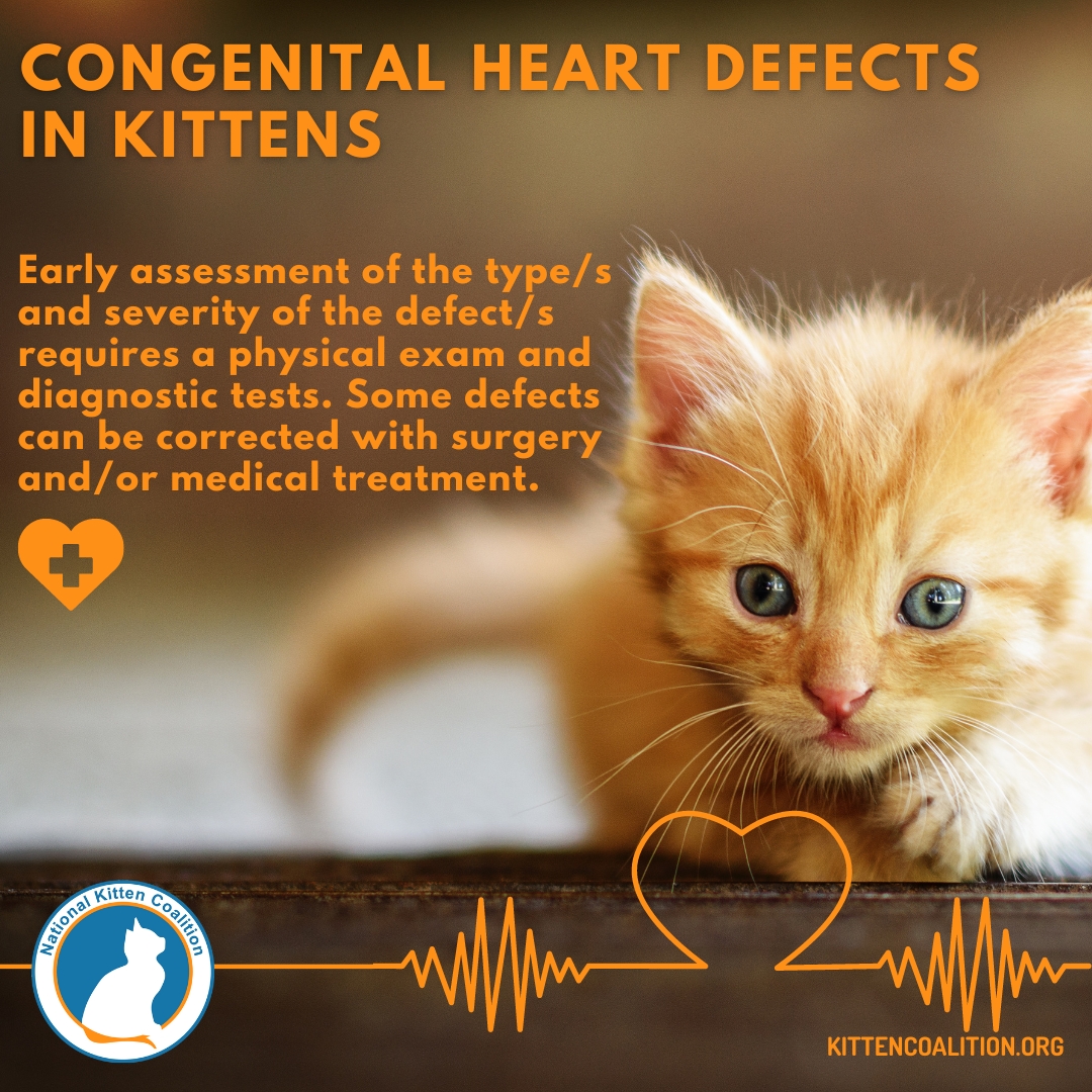
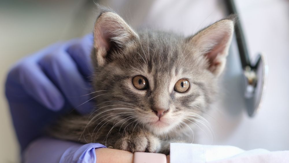
If any of the above symptoms are observed, a veterinarian should be immediately contacted. If recognized early, some defects can be corrected with surgery and/or medical treatment. The veterinarian will perform a physical exam and diagnostic tests to assess the severity of the defect. These tests may include:
- Complete Blood Count (CBC)
- Urinalysis
- Electrolyte panel to measure minerals (g., sodium, calcium and potassium), which are essential for many key bodily functions
- Electrocardiogram (ECG), which records the heart’s electrical activity
- Chest x-rays
- Echocardiogram, an imaging test that uses ultrasound to monitor heart
function
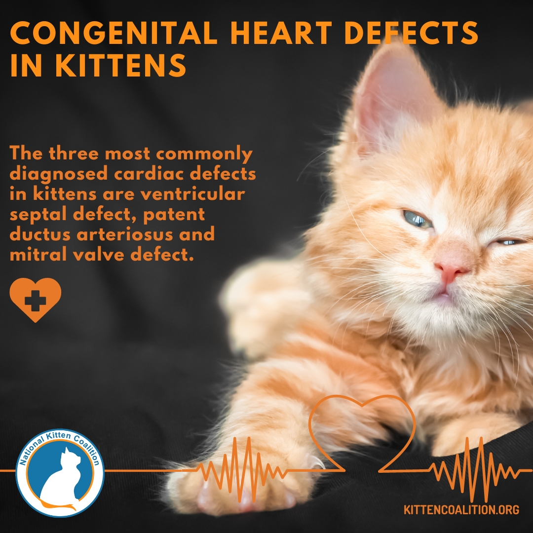
Although cardiac conditions are relatively rare in kittens, the three most commonly diagnosed defects are: ventricular septal defect (VSD), patent ductus arteriosus (PDA) and mitral valve defect (MVD)
- VSD occurs when there is a hole between the left and right ventricle chambers. If the hole is small, a kitten may be asymptomatic and can often have a normal life expectancy. With medium and large holes, symptoms become more apparent, and life expectancy decreases.
- PDA occurs when a fetus’s temporary blood vessel that connects the aorta and pulmonary artery fails to close within days of birth. This causes too much blood to flow from the heart to the lungs and can cause heart failure. A veterinarian can repair the defect with a surgery that ties off the vessel with either sutures or clips. This can be done when the kitten is a few months old, or earlier if the kitten is deemed physically able to tolerate the surgery.
- Mitral valve defects are malformations of the valves between the atria and ventricles, which should close to prevent backflow of blood when the heart beats. Mildly affected cats may live for years without any signs, but the outlook is poor for severely affected cats. Symptoms include difficulty breathing, reduced appetite and thirst and possible arrhythmias (irregular or abnormal heartbeats).
Less common congenital heart defects include:
- Atrial septal defect (ASD), which is a hole in the septum, the muscular bands that separate the right and left atria. The prognosis is often guarded to poor and depends on the size and location of the defect, along with concurrent abnormalities.
- Tricuspid valve defect (TVD) leads to blood being regurgitated back into the right atrium, which leads to enlargement of the right atrium and ventricle, and, in severe cases, to right heart failure. Symptoms include fatigue, tachypnea (increased rate of breathing) and ascites (accumulation of fluid in the abdomen).
- Endocardial fibroelastosis causes the walls of the left atrium, ventricle and area around the mitral valve to become enlarged and thickened. This is a hereditary defect seen most commonly in Siamese and Burmese cats. Symptoms of heart failure will usually appear before the kitten reaches 6 months of age.
- Aortic and pulmonic stenoses involve the narrowing of a blood vessel or valve. This causes blood flow to be reduced. Aortic stenosis is the narrowing of the aortic valve, which causes the left ventricle to work harder to pump blood into the aorta. This additional strain can result in heart failure and/or sudden death. In mild cases, treatment is often not required; however, in moderate to severe cases, long-term medication may be necessary. Pulmonic stenosis causes obstruction of blood flow from the right ventricle through the pulmonary valve. Although surgery and medications may help in certain instances, the outlook is poor if atrial fibrillation or right-sided congestive heart failure is present.
- Tetralogy of Fallot consists of four physical abnormalities, including: a ventricular septal defect (a hole between the two ventricles); pulmonic stenosis; an overriding aorta (varying degrees of the aorta rotating to the right); and right ventricular hypertrophy (thickening of the muscle fibers of the right ventricle). The outlook for survival is guarded, but some cats may survive into adulthood depending on the severity and size of the abnormalities.
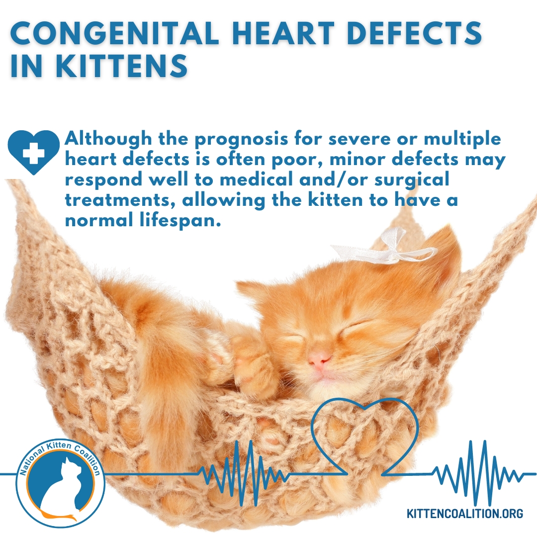
Fortunately, the frequency of congenital heart defects in cats is estimated to be less than 1-2%. Although the prognosis for severe or multiple heart defects is often poor, minor defects may respond well to medical and/or surgical treatments, allowing a kitten to have a normal lifespan.